Niosomes: Classification, preparation and application
Abstract
Vesicular medication delivery system, for instance, niosomes may be a novel medication delivery system, during which the answer is enclosed in vesicle which is formed by Non-ionic surfactant. Structurally, niosomes are almost like liposomes, therein they're also made from a bilayer. However, the bilayer within the case of niosomes is formed from non-ionic surface actives agents instead of phospholipids as seen just in case of liposomes. Niosomes tackled the difficulty of insolubility, instability, low bioavailability and fast debasement of medicines. The both hydrophilic or lipophilic nature (amphiphilic nature) of niosomes increase their capacity in encapsulating lipophilic or hydrophilic drugs.Different type additives, such as cholesterol, are often wont to maintain the rigidity of the niosomes structure. The basic aspects of niosomes, such as their structural components, methods of preparation, and current applications to various diseases.
Keywords
Niosomes, method of preparation, limitations, application
INTRODUCTION
The concept of a drug-delivery system mentioned to a process of administered pharmaceuticals components at a predetermined rate to realize a therapeutic effect in humans or animals at a diseased site, and at an equivalent time, reducing the concentration of the medication in surrounding tissues. When the action of localized drug enhances the efficacy of drugs/substances they reduce systemic toxic effects to tissues 1. Among these systems, liposomes and niosomes are well-documented vesicular drug-delivery systems 2, 3, 4, 5. The niosomesare improved the therapeutic excecution of encapsulated drug molecules by protecting the drug from biological environments, leading in to their delayed clearance 6. The first niosome formulations were developed and patented by L’Oreal in 1975. The properpresents of surfactants and charge inducing agents from the thermodynamically stable vesicles. Niosomes are mostly studied as an alternative to liposomes because they alleviate the disadvantages associated with liposome 7.
SALIENT FEATURES OF NIOSOMES 8, 9, 10
-
Niosomes can entrap solutes.
-
Niosomes are osmotically active and stable.
-
Niosomes have an infra-structure comprising hydrophobic and hydrophilic for the foremost part together thus likewise oblige the medication atoms with a thorough kind of dissolvability.
-
Niosome discharge the medication in a controlled way by means of its bilayer which give supported arrival of the encased medication, so niosomes fill in as medication warehouse in the body.
-
Targeted drug delivery of niosomes as vehicle can likewise be perfects utilizing niosomes the medication is vehicles specifically to the part where the remedial effect is required.
-
They improve the solubility and oral bio availability of poorly soluble drugs and also enhance the skin permeability of drugs when applied topically.
-
Niosomes show flexibility in their structural characteristics and should be designed according to the required situation.
-
Niosomes may be inhensed the performance of the molecules of the drug.
-
Better availability to the actual site, just by protecting the drug from biological environment.
-
Niosomes increase the steadiness of the entrapped drug.
ADVANTAGES OF NEOSOMES 11, 12
-
Bioavailability Improvement
-
Niosomes increase the invigorative performance of the medication particles by postponed relaxation from the diffusion, shielding the medication from natural condition and limiting efficacy to focus on cells.
-
Niosomal dispersion during an aqueous phase are often emulsified during a nonaqueous phase to manage the delivery.
-
Niosomes can increase the rate of drug and administered normal vesicle in external nonaqueous phase.
-
They are osmotically active and stable, also as they increase the steadiness of entrapped drug.
-
They improve oral bioavailability of poorly absorbed drugs and enhance skin penetration of medicine.
STRUCTURE OF NIOSOMES 13, 14, 15
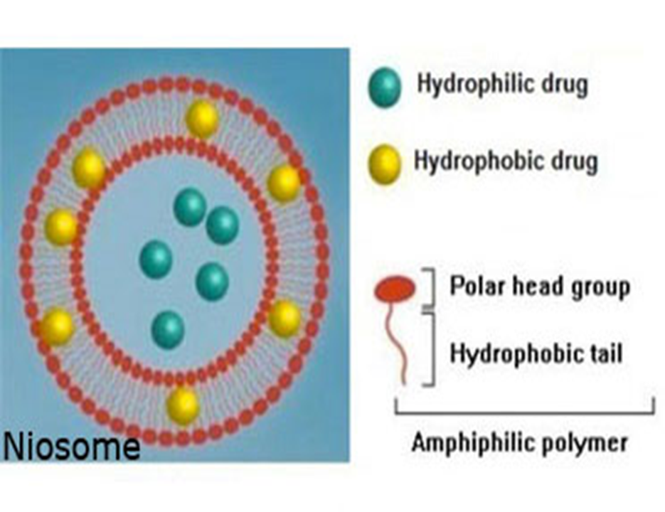
Structurally, niosomes are almost like liposomes, therein they're also made from a bilayer. The bilayer within in the case of niosomes is formed from non-ionic surface actives agents instead of phospholipids as seen within the case of liposomes. Niosomes could also be unilamellar or multilamellar counting on the tactic wont to prepare them. The niosome is formed of a surfactant bilayer with its hydrophilic practicable exposed on the surface and within in the vesicle, while the hydrophobic chains face one another within in the bilayer. Therefore, the vesicle catch hydrophilic drugs within the space enclosed into the vesicle, while hydrophobic drugs are embedded within the bilayer itself. A typical niosome vesicle would contains a vesicle forming ampiphile i.e. a non-ionic surfactant like Span-60, which is typically stabilized by the addition of cholesterol and a little amount of nonionic surfactant like diacetyl phosphate, which also helps in stabilizing the vesicle.
CLASSIFICATION
The three factors of niosomes are classified as a function of the amount of bilayer or as a function of size or as a function of the tactic of preparation. The various types of niosomes are described below:
-
Multi lamellar vesicles
-
Large unilamellar vesicles
-
Small unilamellar vesicles (SUV)
Multilamellar vesicles (MLV)
It consists of a number of bilayers surrounding the aqueous lipid compartment separately. The approximate size of those vesicles is 0.5-10 µm diameter. Multilamellar vesicles are the foremost widely used niosomes. This type of vesicles are highly suitable as drug carrier for lipophilic compounds.
Large unilamellar vesicles (LUV)
Niosomes of this sort have a high aqueous/lipid compartment ratio, in order that larger volumes of bio-active materials are often entrapped with a really economical use of membrane lipids.
Small unilamellar vesicles (SUV)
The approximate size of this vesicle are 10-100 nm and this types of vesicle is prepared from multilamellar vesicles by sonication method, French press extrusion electrostatic stabilization in that the inclusion of diacetyl phosphate in 5-carboxyfluorescein loaded Span based niosomes.16
METHOD OF PREPARATION
A) Hand shaking method (Thin film hydration technique)
The hand shaking method is performed by the combination of vesicles forming ingredients like surfactant and cholesterol are dissolved during a volatile organic solvent in a round bottom flask. The ejection of organic solvent at normal temperature using rotary evaporator leaving a thin layer of solid mixture deposited on the wall of the flask. The drained surfactant film an often rehydrated with aqueous phase at 0-60°C with gentle agitation. This process forms typical multilamellar niosomes 17.
B) Ether injection method
This method provides a way of creating niosomes by slowly introducing an answer of surfactant dissolved in ether into warm water maintained at 60°C. The surfactant mixture in ether is injected through 14-gauge needle into a solution of fabric. Vaporization of ether results in formation of single layered vesicles. Depending upon the conditions used, the diameter of the vesicle range from 50 to 1000nm 18.
C) Extrusion method
In this method, niosomes were prepared using C16G2, a chemically depend non-ionic surfactant by extrusion through a polycarbonate membrane. these studies not only demonstrate the effect of number of extrusions on vesicles size but also the effect of size on encapsulation of drug 17.
D) Reverse phase evaporation technique
In this method, surfactant is dissolved in chloroform and added into the 0.25 ml volume phosphate saline buffer solution is emulsified to get w/o emulsion. The mixture is then solicited and subsequently chloroform is evaporated under reduce pressure. The lipid or surfactant forms a gel first and subsequently hydrates to form vesicles 18, 17.
E) Bubble method
It is modern technique for the one step preparation of niosomes without the use of organic solvents. It consists of round-bottomed flask with three necks placed in water bath to control the temperature. The water-cooling refluence and thermometer is capacity into the first and second neck and nitrogen supply through the third neck. Cholesterol and surfactant are the dispersed together in this buffer (pH7.4) at 70c 6, 18.
F) Sonication
The first time niosomes prepared by Baillie et al. 1986 with the help of sonication method. In this method, surfactant: cholesterol (150 micro.mol.) mixture was dispersed in 2ml aqueous introduce vial. Spread is subjected to probe sonication for the three minutes at six hundred centigrade. These systems confluent the formation of MLVs which are subjected to ultrasonic vibration. Sonicator is two type Probe and Bath sonicator. Probe sonicator is use when sample volume is small size and Bath sonicator is use when sample volume is large 17.
G) Micro fl uidization method 19
This method may be a current strategies to plan unilamellar vesicles of characterized estimate circulation based on submerged jet principle, during this strategy two fluidized streams connect at ultrahigh speeds, in accurately characterized smaller scale channels inside the interaction chamber. The results in more popular consistency, smaller size and better reproducibility of niosomes shape 19.
H) Separation of unentrapped drug 20, 21, 22, 23, 24, 25, 26
The removal of unentrapped solute from the vesicles are often done by various techniques, like dialysis, gel filtration and centrifugation.
1) Dialysis
Dialysis is one among most vital technique used for removal of unentrapped drug from vesicles.
2) Gel Filtration
In the gel filtration systems discarded of unentrapped drug by gel filtration system of niosomal dispersion through a Sephadex – G-50 column and elution with phosphate buffered saline or normal saline.
3) Centrifugation
The niosomal suspension is centrifuged and therefore the supernatant is separated. The small size globules is bleached then resuspended to get a niosomal suspension free from unentrapped drug 25, 27.
APPLICATIONS OF NIOSOMES 28, 29
Niosomesare used for studying the character of the immune reaction provoked byantigens.
-
It is used as drug Targeting.
-
It is used as Anti-neoplastic Treatment i.e. Cancer Disease.
-
It is used as Leishmaniasis i.e. Dermal and Mucocutaneous infections e.g. Sodium stibogluconate.
-
Niosomes as carriers for Hemoglobin.
-
It is more suitable for delivery of peptide drugs.
-
Niosomes is best used as a carrier for hemoglobin.
-
It is used in Studying Immune Response.
-
Transdermal drug delivery systems utilizing niosomes.
-
It is used in ophthalmic drug delivery.
-
Niosomal system are more helpful for diagnostic agents.
CONCLUSION
Niosomes represent a promising drug delivery module. They present a structure almost like liposome and hence they're going to represent alternative vesicular systems with regard to liposomes, thanks to the niosome ability to encapsulate different sort of drugs within their multi environmental structure. Niosomal drug delivery system is one is the best suitable examples of great evolution in drug delivery technologies and nanotechnology. The ionic drug carriers are comparatively toxic and unsuitable. Whereas niosomal carriers more time safer and effective. And also handling and storage of niosomes require no special condition. Various sort of drug deliveries are often possible using niosomes like targeting, ophthalmic, topical and parenteral.
AUTHOR CONTRIBUTION
All authors contributed equally.